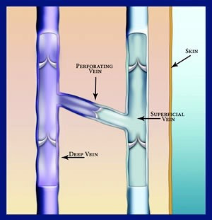
Superficial veins are located just under the surface of the skin. The great and small saphenous veins are the main superficial leg veins.
The great saphenous runs down the inner thigh and leg. The small saphenous begins behind the knee and runs down the back of calf. Both of these veins branch out into a huge network of smaller superficial veins.
In healthy veins, blood flows from superficial veins and their branches through perforator veins and on to deep veins. When valves in superficial veins don’t work properly, varicose veins develop. These can be treated with radiofrequency ablation (RFA) and phlebectomy. The problem superficial vein is closed and other healthy veins take over.
Deep veins are large veins that pump blood up to the heart. They are located in the muscle and can only be seen via ultrasound. With every step you take, the calf muscles compress deep veins to force blood toward the heart.
When deep vein valves fail, deep vein thrombosis (DVT) can occur. Problem deep veins can be improved with compression therapy and by treating superficial veins.
Perforating veins — or perforators — carry blood from the superficial veins to the deep veins. When the valves of perforator veins don’t work properly, blood is pushed back into the superficial veins. This worsens existing varicose veins and can lead to skin changes and the development of venous ulcers.
Perforating veins can be treated with endovenous laser treatment (EVLA). The problematic vein is sealed shut and other healthy veins take over.

While problems with superficial veins are visible to the eye, there could be much more going on that you cannot see. With ultrasound imaging, Dr. Bellamah can view the entire network of diseased veins. Finding the deeper source of visible varicose veins is important for developing an effective treatment plan.